
Call Us:
+91 7304075735
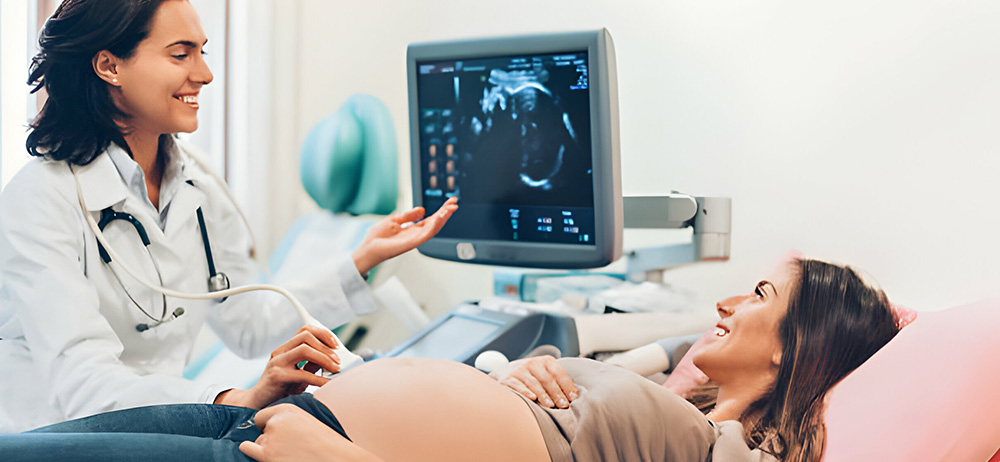
Ultrasound sonography, often referred to simply as ultrasound, is a non-invasive medical imaging technique that uses high-frequency sound waves to create real-time images of the inside of the body. It is a versatile and widely used diagnostic tool, providing valuable insights for various medical purposes. Here's an overview of ultrasound sonography
Ultrasound works on the principle of sending sound waves into the body and detecting the echoes produced as the waves bounce off internal structures. These echoes are then translated into visual images.
Ultrasound is a non-invasive and painless procedure. Depending on the type of examination, preparation requirements may vary. For abdominal scans, fasting may be necessary, while pelvic or obstetric ultrasounds may require a full bladder for optimal imaging.
Ultrasound sonography stands as a cornerstone in modern medical diagnostics, offering a safe and efficient means of visualizing internal structures for accurate diagnosis and treatment planning. Its widespread use across medical specialties underscores its importance in providing valuable medical insights with minimal invasiveness.
For more information please contact Laxmi Advanced Womencare @ +91-7304075735 / charmithakker@yahoo.com

